Whole-slide imaging is a qualified alternative to traditional microscopy. With approximately 1,000 pathology labs around the world converting to digital pathology systems, the accuracy of digital pathology images compared to glass slides is no longer an obstacle.
Modern dermatologists are turning to whole-slide imaging to take advantage of remote access, portability, ease of sharing, virtual archiving, and user-friendly tools. By examining the concordance with glass slides, technology training, and intraobserver variability, we will demonstrate why clinics should embrace laboratories that have undergone a digital transformation.
Digital slides are reliable and accessible.
Ensuring every patient receives the highest quality of care means dermatologists require complete trust in the results of their reads. With diagnosis from digital slides equivalent to diagnosis from glass slides, digital pathology is a proficient, high-tech replacement for optical microscopes.
Surprisingly, less than 5 percent of global pathology sites use digitalized workflows to handle all of their primary diagnoses. Misconceptions about the barriers to entry—including cost, labor, software, and memory—prevent dermatologists from moving to whole-slide imaging. However, partners like PathologyWatch remove these barriers because they can, in the context of providing tissue processing and/or interpretation, carry the burden of cost and provide the necessary hardware, implementation, labor to digitize, and software maintenance to make it simple for clinics to access electronic records.
The only remaining hurdle is familiarity, which is essential to building confidence in any new form of technology. In a study from the Department of Pathology, University of Pittsburgh Medical Center, pathologists who received proper training interpreted digital imaging with 16 percent greater accuracy than those without training.
Digital pathology provides significant clinical benefits.
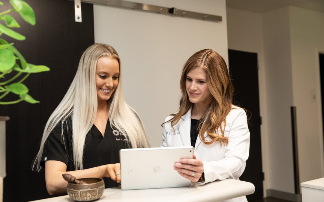
Incorporating whole-slide imaging into your practice means providing patients with industry-leading, state-of-the-art processes. “When patients come to my office I can show them the whole slide image on the computer screen, point out things of significance, and tell them what they mean,” says Sylvia Asa, MD, PhD. “Sitting with patients and reviewing their pathology is a critical element that brings us to the patient and makes patients understand the importance of what we do. And that has always been a big challenge for pathology.”
Intraobserver variability is a chance for collaboration.
Because glass slides and digital pathology images are equivalent, a challenging read on a digital device will result in the same challenges when analyzed through a microscope. For this reason, it is expected that the same dermatologists or pathologists will render a different diagnosis on a certain percentage of cases depending on the day or modality on which they are viewed.
However, digital imaging offers dermatologists an advantage when it comes to challenging reads. Unlike glass slides, specimens on a digital image viewer* can be simultaneously viewed and annotated by colleagues around the world at a moment’s notice. With the demand for pathologists in the US expected to increase 16 percent by 2030, adapting to the efficiency and collaborative offerings of digital imaging can keep your practice up to speed with a higher workload.
Aligning with a laboratory like PathologyWatch will help your dermatology practice access the performance and productivity of whole-slide imaging. By understanding the concordance between digital pathology images and traditional microscopy, the value of appropriate training, and intraobserver variability in analog and digital reads, you have the assurance that digital pathology is right for your practice.
*Images shown are not intended to be used for the diagnosis or treatment of a disease or condition.