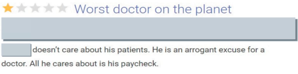
Dan Lambert Shares How the Pandemic Has Upset Healthcare and Five Ways to Fix It

In a new article published by Forbes, Dan Lambert, CEO of PathologyWatch, examines how the pandemic is causing harm to particular functions of the healthcare industry.

While COVID-19 will have a long-lasting physical and economic impact worldwide, it has also exposed some troubling problems within the healthcare community. These include steep financial challenges facing hospitals and private practices, patients delaying medical procedures, overlooking pre-existing healthcare industry concerns, and bottlenecks in pathology lab workflow.

Dan explains, “When a pathology lab delays processing skin specimens or other biopsies to focus on COVID-19 cases, it unintentionally increases the patient’s level of risk.” He adds, “Some estimates claim these coronavirus postponements will lead to over 10,000 cancer fatalities in the next decade. By reducing the wait time for day-to-day lab results today, we can save lives tomorrow.”

Dan also shares five action items that could help the healthcare industry improve the situation before it’s too late. These include telehealth adoption, hospital bailouts, digital data transfer, alternative insurance options, and academic standards.

To read the full Forbes article, click here.

Diagnostic Markers for Melanocytic Lesions

Melanocytic lesions, commonly found in 23 percent of skin biopsies, can sometimes be challenging to diagnose based on morphologic criteria alone. However, utilizing diagnostic markers can make a significant difference in the accuracy of a melanocytic lesion diagnosis. 

As a dermatologist, you rely on your partnering dermatopathology laboratory to demonstrate expertise when it comes to diagnosing your melanocytic lesion cases. By understanding why diagnostic markers are ordered and examining the more prevalent types of immunohistochemical stains, you can play a greater role in providing your patients with optimal care.

Why are diagnostic markers ordered?

In a survey of 160 dermatopathologists, 99 percent indicated they ordered additional immunohistochemistry (IHC) or molecular tests out of concern for patient safety. Because melanocytic lesions can produce harmful results if a melanoma diagnosis is missed, laboratories often order diagnostic markers to make sure the results are as definitive as possible, utilizing the most reliable tests.

Medicare has released guidelines to cut costs, reduce overutilization, and ensure that orders for diagnostic markers meet medically necessary criteria. These directives indicate that “a pathologist must first review the H&E [hematoxylin and eosin] stain prior to ordering special stains or IHC.” They add that “the medical necessity for the special stain . . . must be documented in the surgical pathology report.”

The most proficient and ethical dermatopathologists avoid overbilling by ordering additional stains only when they are truly necessary. 

What are the different types of markers?

Immunohistochemical stains are used to help pathologists arrive at an accurate diagnosis. As a dermatologist, it’s essential to be familiar with the stains as they will eventually end up in your report.

These antibodies are the most familiar diagnostic markers: 

  • S100 – The S100 protein is found in 95 percent of primary cutaneous melanomas, making it a popular stain. Diagnostic challenges can occur if the tissue was formerly frozen, had a low fixation time, or underwent enzymatic pretreatment with trypsin. S100 protein detects melanocytes, as well as neoplasms of the nerve sheath such as schwannomas.
  • HMB45 Human Melanoma Black-45 was derived from extracts of pigmented melanoma in lymph nodes. HMB-45 antibody is specific for melanocytes and exposes patterns of nevi maturation. It can also detect patchy gp100 patterns in primary cutaneous melanomas.
  • MART1Melanoma Antigen Recognized by T Cells 1 identifies most melanocytic lesions. It is a cytoplasmic stain and can highlight the dendritic cell processes of melanocytes. It is also known as Melan A.

These markers have been discovered recently:

  • MiTF – Microphthalmia Transcription Factor is a sensitive and specific marker for melanocytes. MiTF has superior sensitivity and specificity to S100 and HMB45. This is a nuclear stain and can be used to quantify pagetoid spread of melanocytes. However, other cells can express MiTF, such as macrophages, smooth muscle cells, and fibroblasts.
  • SOX10  SRY-Related HMG-Box Gene 10 is a nuclear transcription factor seen in melanocytes with a similar staining pattern as MiTF. SOX10 is also seen in nerve sheath tumors. 
  • PRAME – Preferentially Expressed Antigen in Melanoma is a tumor-associated antigen expressed in cutaneous melanoma, ocular melanoma, and other nonmelanocytic malignant neoplasms such as lung and breast carcinoma. The PRAME immunohistochemical stain has been shown to be diffusely positive in the nuclei of most melanoma cells and negative in most benign nevi. In addition, its specificity for malignant cells has made it a candidate for targeted immunotherapy.

Because the diagnosis of melanoma can sometimes be difficult, additional stains or second opinions are sometimes utilized. 

A 2019 pathology study indicates “second opinions rendered by dermatopathologists improve reliability of melanocytic lesion diagnosis.” Utilizing digital slides from full-service dermpath labs like PathologyWatch enables multiple experts to collaborate on difficult cases simultaneously. When a dermatopathology group includes expert dermatopathologists that are trained and accustomed to a standardized laboratory process, there is no need to duplicate any ancillary staining, thereby reducing costs. Furthermore, seeking second opinions earlier in the process can help limit the number of stains required for definitive diagnosis. 

Performing biopsies on melanocytic lesions will be a constant cause of concern for patients and their dermatologists. With the high stakes of melanoma and the possibilities of missed diagnoses, it is critical to know why diagnostic markers are ordered while becoming familiar with the most popular immunohistochemical stains to keep your patients safe. 

Molecular Testing in Melanoma 

Dermatologists understand the concern and danger patients face when biopsies reveal melanoma. Since almost 7,000 Americans are predicted to die from melanoma in 2020, and some therapeutic regimens for advanced disease are contingent upon molecular tumor markers, clinics must take an active role in accelerating the molecular testing process.

Dermatologists can benefit from a baseline understanding of molecular testing. By identifying why molecular tests are ordered, exploring the most common forms of testing, and examining the testing workflow, dermatologists can help to ensure their patients receive life-saving care.

Why are molecular tests ordered?

When a diagnosis of advanced melanoma occurs, molecular diagnostic testing is critical to revealing opportunities for targeted therapies and immunotherapies. Targeted therapies treat melanoma by inhibiting genetic driver mutations. One of the most commonly mutated genes in melanoma is BRAF, which affects about 50 percent of all melanoma cases. Mutations in the BRAF gene produce a protein that can cause cancerous cells to grow and divide. In addition, tumors with certain BRAF mutations are sensitive to targeted therapy with BRAF and MEK inhibitors, which is associated with prolonged survival. Another commonly mutated gene in melanoma is NRAS, which can also be targeted by MEK inhibitors.

Melanomas with mutations in the C-KIT gene are less common but can also be tested for. Typically, C-KIT melanomas occur on the palms, hands, feet, or mucosal areas like the mouth. Some targeted therapies, such as imatinib, can be utilized in tumors with C-KIT mutations.

Immunotherapy utilizes the body’s own immune system to treat melanoma tumors. A biomarker called tumor mutational burden has been associated with response to immunotherapies. Tumor mutational burden can be determined by certain types of molecular testing.

Molecular testing is recommended for at-risk patients with stage 3 and stage 4 metastatic melanoma. While stage 3 can suggest satellite tumors or multiple lymph nodes matted together, stage 4 indicates cancer has spread to other areas of the body, such as the liver, soft tissue, spinal cord, or lungs. A patient in either stage can expect surgery, targeted therapy and/or immunotherapy, and possible radiation treatment.

What are the most common forms of testing?

When testing detects cutaneous malignant melanoma in the early localized stage, patients have a five-year survival rate of 99 percent. However, a belated diagnosis that results in cancer spreading to regional or distant locations can reduce the five-year survival rate to as little as 25 percent.

Molecular testing is most commonly performed on a sample of the patient’s melanoma tumor. Testing procedures are often determined by what is covered by the patient’s insurance. Next-Generation Sequencing (NGS) performs full characterization and provides the most genetic information. This highly sensitive test can identify mutations in melanoma tumor tissue simultaneously in BRAF and hundreds of other genes, including NRAS and C-KIT. Some NGS tests can also measure tumor mutational burden, a biomarker for response to immunotherapy.

Real-time polymerase chain reaction (RT-PCR) is another form of molecular testing that detects the most common mutations (hot spot mutations) in a gene. While PCR testing specializes in low numbers of gene targets, it can offer faster turnaround time for results when only one or a few genes are of interest. NGS testing can simultaneously detect mutations across thousands of target regions. Either of these genetic tests is optimal for BRAF mutation analysis.

What is the molecular testing workflow?

Traditionally, oncologists handle molecular testing. However, dermatologists can speed up the process by requesting the tests from their dermatopathology lab partner, which should have protocols to manage and report mutation tests. Experienced full-service dermatopathology labs like PathologyWatch stay up to date on the latest molecular testing processes. In addition to managing tests for dermatology clinics, the academic-level dermatopathologists at PathologyWatch also manage testing for oncologists.

Once the laboratory makes the melanoma diagnosis, EMR integration is a reliable way to share fast and accurate results with the dermatologist, who is an integral part of the patient’s care team. After receiving the results, the dermatologist can refer the patient to an oncologist, who will start life-saving treatment based on the molecular testing results.

Though representing only 1 percent of all skin cancers, melanoma’s ability to spread to other parts of the body makes it highly lethal. As a dermatologist, knowing why molecular tests are ordered, understanding the different types of testing, and being aware of the testing workflow can help you get your patients the prompt treatment they require.

PathologyWatch Announces Locum Tenens Dermatopathology Services

Full-service lab PathologyWatch now provides remote, academic-level dermatopathology services to laboratories in need of temporary or long-term support.

Digital dermatopathology service provider PathologyWatch is now offering top-tier dermatopathology services to laboratories in need of temporary or long-term locum tenens support. Utilizing the advantages of digital pathology, PathologyWatch provides instant remote access to a workforce of academic-level dermatopathologists.

Commonly, a laboratory in need of locum tenens assistance is responsible for providing travel arrangements, lodging and per diem to attract qualified talent. Using an integrated digital pathology system, PathologyWatch eliminates costly overhead by offering a network of expert dermatopathologists available 24/7 for consultation. As a result, laboratories find a seamless transition to help them to continue running at full capacity.

“Providing locum tenens support to companies experiencing overflow due to staffing or seasonal variables is the next logical step for PathologyWatch,” says CEO and co-founder Dan Lambert. “Our locums tenens support is ideal for pathology labs and dermatology clinics that have a limited number of in-house dermatopathologists. The on-demand services require no minimum or advanced notice. When dermatopathologists have an influx of samples, have a life event, or even want to take a vacation, they can simply turn on the services for as long as they need them.”

In addition to a significant reduction in travel costs, PathologyWatch provides labs with 24/7 digital access to all pathology slides without purchasing a scanner. This also includes improved access to subspecialists and second opinions. Meanwhile, extensive insurance coverage means PathologyWatch can accommodate the majority of laboratory customers nationwide.

“Using PathologyWatch for locums coverage has given me access to high-quality dermatopathologists when I needed the coverage with ease and flexibility,” said Emily Green, dermatopathology lab director at Golden State Dermatology. “I highly recommend their services.”

For more information on PathologyWatch, please visit PathologyWatch.com. PathologyWatch follows all state and federal regulations. As such, the specific coverage arrangement may vary from state to state.

About PathologyWatch

PathologyWatch is the groundbreaking leader of digital dermatopathology services. Through these services, dermatology clinics, hospitals and laboratories can improve operational efficiency by speeding up workflow and enhance patient outcomes by utilizing the PathologyWatch expert professional team and partner laboratory services. This can facilitate best-in-class reads and, in some cases, enable additional revenue to the practice by in-housing pathology. With an intuitive and easy-to-implement digital pathology solution that includes access to top-tier dermatopathologists and a streamlined clinical workflow that interfaces directly into the EMR, PathologyWatch brilliantly combines state-of-the-art technology and clinical decision making to deliver unprecedented patient care.

3 Ways Digital Pathology Is Instrumental in the Shared Decision-Making Process

Sometimes, seeing is believing.

This is never more true than when discussing a treatment plan for a medical condition. “Communicating the importance of medical evidence and a balanced representation of options is the first step toward accelerating patient engagement in shared decision making,” concluded the groundbreaking discussion paper Communicating with Patients on Health Care Evidence.

This research found that 80 percent of patients want their provider to tell them the full truth about their diagnosis even though it may be unpleasant. Around 70 percent of people surveyed want their provider to share the risks associated with each option. But how can a healthcare provider share clear, definitive information for their patients who are often unfamiliar with terminology and find it difficult to fully comprehend the ramifications of the diagnosis? 

The answer is simple: Provide a visual aid. 

The aforementioned study published by the Institute of Medicine of the National Academies examined the effects of using visual aids as part of the patient’s decision-making on treatment plan options. Researchers found that when patients are given decision aids—such as educational booklets, DVDs, or interactive tools—to help them make treatment choices, they are more knowledgeable and satisfied with their care. 

If visual aids prove beneficial for patients to better understand their diagnoses and the necessary treatment plan, then let’s discuss the three exciting ways digital pathology acts as an effective decision aid for patient care and heightens the quality of care delivered. 

1. Encourages Patient Engagement

At PathologyWatch, we utilize the collaborative capabilities of digital technology as it provides full access to a patient’s test slide through our EMR. And that information can also be shared with a patient to assist in the decision-making process for designing a treatment plan. 

In many cases, patients are better able to understand their diagnoses when they can see the pathology associated with their cases. This isn’t just about delivering information to a patient. Instead, as our dermatology clients attest, it invites a personal conversation that centers around a patient’s concerns and goals for treatment. 

Typically practiced in breast cancer cases, for example, shared decision-making (SDM) provides a collaborative strategy for a patient and the healthcare provider to discuss medical options. Most consumers (95 percent) believe it is important that doctors tell them about the results of medical research when making treatment decisions. By pairing the patient’s values and priorities with the physician’s expertise, both parties can talk about the best possible option.

2. Provides Faster Results

Experts predict that the global number of new cancer cases will rise by 70 percent over the next two decades. This means pathology labs will need to find ways to accommodate the increase in demand through cost-saving efficiency and diagnoses. 

“Digitizing your workflow is not simply a question of upgrading your lab’s hardware and IT,” says Ivo Van den Berghe, MD. “It’s also about rethinking your ways of working. . . . Digital pathology replaces the subjective nature of manual slide inspection under the microscope.”

Digital transformation also creates more efficiency. “In the old situation, a lab assistant would have to manually sort and archive 300 tissue samples each day,” Van den Berghe says. “Now the samples are sorted and archived automatically. This saves our lab assistants a lot of time.” Such expediency means doctors can meet with their patients sooner, armed with visual test results, and discuss more timely treatment plan options that can improve the recovery rate. 

3. Provides a Comprehensive Perspective

In cases where the diagnosis involves a rare condition, digital pathology can make it possible for doctors to tap into expertise available throughout the world for a comprehensive understanding of an irregular case. “We’re connecting with laboratories worldwide to get a better understanding of rare tumors and how to treat them,” Van den Berghe says. “Digital pathology enhances clinical confidence in our findings by delivering the right result the first time.”

Patients trust their doctor to deliver accurate results. With digital pathology, they can get their results quickly, participate in shared medical decisions, and potentially benefit from a more comprehensive perspective of their diagnosis and ideal treatment options.