Journal of Pathology Informatics: High-Fidelity Detection, Subtyping, and Localization of Five Skin Neoplasms Using Supervised and Semisupervised Learning

Pathologywatch Implements AI Technology That Detects 5 Skin Lesion Types with Higher Sensitivity than Previously Published AI Model

Authors: James Requa, Tuatini Godard, Rajni Mandal, Bonnie Balzer, Darren Whittemore, Eva George, Frenalyn Barcelona, Chalette Lambert, Jonathan Lee, Allison Lambert, April Larson, Gregory Osmond

Highlights

  • Including a large dataset of hand-drawn annotations increases the sensitivity of AI.
  • Integrated supervised and semisupervised learning is superior to other AI models. 
  • Multilevel training permits subtyping, localization, and margin detection.
  • Dermpath involvement in model development can ultimately improve model performance.
  • Increased confidence in AI-assisted diagnoses can lead to improved patient care.

Abstract

Background

Skin cancer is the most common malignancy diagnosed worldwide, accounting for over 30% of all cancer diagnoses. Equally troublesome is the 77% increase in nonmelanoma skin cancers over the last several decades and a 250% increase in melanoma cases in children, adolescents, and young adults since 1973. 

Objective

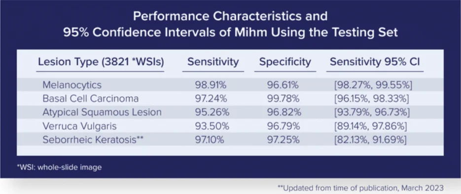
The goal of this study was to create an AI model that detects and classifies skin lesions with a higher degree of sensitivity than previously demonstrated. This AI model (i.e., Mihm) has the potential to match and eventually surpass expert pathologists and improve clinical workflows.

Methods

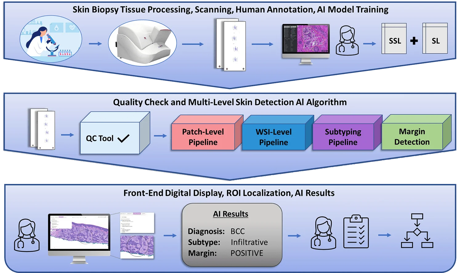
We produced an end-to-end multilevel skin detection system by combining supervised learning (SL) with semisupervised learning (SSL). This system detects five main types of skin lesions with high sensitivity and specificity. In addition, it detects subtypes while also localizing and providing margin status to evaluate the proximity of the lesion to nonepidermal margins. 

Supervised Training Subset

  • 2,188 random WSIs collected by PathologyWatch laboratory between 2013 and 2018
  • Weekly supervised subset consisting of 5,161 WSIs from daily case specimens

Validation Set

  • 250 curated daily case WSIs from PathologyWatch’s tissue archives
  • Included 50 “mimickers”

Testing Set

  • Composed of noncurated daily case specimens collected from PathologyWatch labs between July 20, 2021, and August 20, 2021

Results

By running the testing set through the Mihm Evaluation Pipeline, we could assess the performance characteristics of our AI model. 

PathologyWatch AI

Our multilevel detection algorithm (patch-level, ROI-level, WSI-level) can subtype lesions with an AI overlay in the front-end digital display that localizes diagnostic ROIs and reports on margin status by detecting overlap between lesions and nonepidermal tissue margins.

Conclusion

This AI model, which is the result of collaboration among a team of dermatopathologists, detects five skin lesion types with higher sensitivity than previous AI models. It also provides end users with valuable insights in subtyping, localization, and margin status in a front-end digital display.

This end-to-end system can improve pathology workflows by doing the following:

  • Increasing diagnostic accuracy
  • Expediting the course of patient care
  • Improving patient outcomes

PathologyWatch Raises $25M to Advance AI-Driven Skincare Research and Diagnostics

The Series B funding round will help PathologyWatch digitize biopsies, increase access to top skincare physicians and further AI research.

SALT LAKE CITY, November 16, 2021—PathologyWatch, the leading digital lab and pathology platform for dermatologists, announced today that it has raised $25M in Series B financing with participation from Ceros Capital Markets, Rock Creek Capital, SpringTide, Spark Growth Ventures, Blueprint Health, Blackbrook Management Group and existing investors.

With the new funding, PathologyWatch will broaden its outreach to dermatologists and conduct further research into skincare diagnostics and AI – allowing patients to receive faster diagnoses and more equitable access to dermatopathology services while enabling labs to work more efficiently and cost-effectively. Additionally, the funding will support operations as the business expands across the country, adding new labs in Texas, Florida and Arizona.

“We are honored that of all the digital dermpath labs available, dermatologists are increasingly choosing to partner with us,” said Dan Lambert, PathologyWatch CEO and cofounder. “As digital dermatopathology continues to grow, it has the potential to reduce the costs of pathology by billions while creating better patient care for everyone across the country. We’re thrilled to be winning so much volume so quickly.”

PathologyWatch provides fully interfaced EMR reporting and 24/7 access to digital slides for dermatology clinics. For their clients, this means more-efficient workflows and direct access to leading dermatopathologists across the country, with broad insurance coverage. PathologyWatch has successfully integrated laboratory information systems, scanners, digital viewer technology and EMRs into an end-to-end solution for dermatologists.

“PathologyWatch has been disruptive to the market and is digitally transforming the industry in unprecedented ways,” said Mark Goldwasser, CEO of Ceros. “With the need for remote health services during COVID, the distributed network of top-tier dermatopathologists alongside a digital viewer that can be accessed anytime and anywhere could not have come at a more fortuitous time.”

“For a dermatology practice to send nearly all skin biopsy volume for cancer diagnosis to PathologyWatch is a no-brainer,” said Austin Walters, founder and managing partner at SpringTide. “The company has worked hard to create a service that outperforms every other from both cost and quality perspectives.”

“Dan is a driven entrepreneur with social good in mind,” said Ryan Brooks, principal at Blackbrook Management Group. “We are part of a cause, not just an investment capital endeavor. In the past, I have been a patient waiting for pathology to come back. When you are waiting for results that could change your life dramatically, you want doctors and tech you can depend on. PathologyWatch will enhance the human experience and enable dermatopathologists to do a better job.”

“I am proud to support PathologyWatch’s mission to provide premier, accessible and affordable digital pathology services to the world,” said Rick Stratford, managing director of Rock Creek Capital. “The ability to provide top-tier pathology services to all communities regardless of location brings hope to patients in underserved areas and can save countless lives. PathologyWatch and other digital platforms like it are democratizing healthcare services and bringing hope and change to our healthcare systems.”

For more information about PathologyWatch, visit pathologywatch.com or contact [email protected].

About PathologyWatch
PathologyWatch is the groundbreaking leader of digital dermatopathology services. Through these services, dermatology clinics, hospitals and laboratories can improve operational efficiency by speeding up workflow and enhancing patient outcomes by utilizing the PathologyWatch expert professional team and in-house lab services. With an intuitive and easy-to-implement digital pathology solution that includes access to top-tier dermatopathologists and a streamlined clinical workflow that interfaces directly into the EMR, PathologyWatch brilliantly combines state-of-the-art technology and clinical decision-making to deliver unprecedented patient care.

Is Melanoma “Overdiagnosed?”

By Darren Whittemore, DO

As one of the most feared types of cancer, melanoma accounts for about 1 percent of skin cancers while contributing to over 7,000 deaths in the United States every year. And those numbers are steadily increasing, with annual incidences rising up to 4–6 percent over the last several decades.

In the age of improved preventative measures, early detection, digital technologies, and better treatments, why are cases of melanoma continuing to rise? Is it our propensity for skin cancer? Or could it be the diagnosis? Are patients receiving the most accurate and reliable information possible? Some voices in the dermatopathology industry believe the pursuit of a “definitive” diagnosis—bolstered with advancements in digital pathology—sometimes leads to biopsies that are collected and tested too soon and ultimately come back with a negative result for malignancy. 

However, others believe it is better to risk overdiagnosis than to miss cases before it is too late. As the industry-wide debate persists concerning melanoma diagnostic processes, let’s discuss points that support both sides of the argument. 

Yes: There are indicators of melanoma overdiagnosis.

“From an epidemiologic perspective, the sharp rise in the incidence of melanoma in the face of stable mortality for the past 40 years [signifies] the epidemiologic signature of overdiagnosis,” says Jason B. Lee, MD, in an article for the American Academy Dermatology Association

Lee echoed some of the concerns expressed by H. Gilbert Welch and his team’s research, which postulates that “the rapid rise in the incidence of melanoma is not due to a true rise in incidence, but it is the byproduct of increased scrutiny, which they refer to as the epidemic of inspection, surveillance, and biopsy of pigmented skin lesions.” 

Welch’s team asserts that the rapid rise in the incidence of melanoma is due to the increasing skin cancer screening activities, low threshold to biopsy, and low threshold to diagnose melanoma by dermatopathologists fueled by heightened public awareness of melanoma, financial incentive, and fear of missing melanoma that has resulted in what they refer to as the “cycle of melanoma overdiagnosis.” 

No: Testing for early detection—even if tests don’t result in malignancy—is good.

Opponents of this overdiagnosing argument—particularly Sancy A. Leachman, MD, PhD, and John D. Gray, an endowed chair in melanoma research—caution against rejecting new technologies for fear of diagnoses not resulting in malignant melanoma:

They suggest that we revert to not biopsying lesions less than 6 mm, rather than making an effort to better understand the biology (and clinical signs) that makes some 2 mm lesions deadly. Shouldn’t our call-to-action be to improve, rather than to decrease, our diagnostic scrutiny? Wouldn’t it be better to utilize the COVID “experiment of nature” to evaluate the true (data-based) impact of decreased melanoma screening, rather than keep all screening programs closed? . . . Should the SPOT Skin Cancer™ screening program be halted completely, as they suggest, or should we purposefully stratify risk and screen those with highest need?

The drive toward innovations in digital pathology and AI technology continues to improve the accuracy of tissue testing. Advances in digital pathology transform our view of the early stages of cellular behaviors. 

But how are we best utilizing this information? At PathologyWatch, we combine state-of-the-art technology and clinical decision-making to deliver optimal patient care with accurate diagnosis. Our clinical team carries extensive experience in identifying and predicting the cellular behaviors of chronic conditions and skin cancers. One of the advantages of having access to this expertise is that dermatologists can collaborate with our clinical team to discuss those factors should there be concerns about early detection. 

Call us to discuss the innovations in digital pathology that can help your practice diagnose with confidence.

Is Your Dermlab a Good Fit? 5 Questions to Ask a Dermatopathology Lab

5 questions to ask your pathology lab
are staff board certified
focused on the client
whole-slide image experience
promote community specialty
prioritize confidentiality
contact pathologywatch today
Are you searching for a new dermatopathology lab? With the onset of digital pathology, finding a lab that is not only equipped with this innovation but also qualified to process whole-slide images swiftly yet accurately may take some time. 

An NIH study found that three out of four respondents agreed that accurate diagnoses can be made with this technology, and over half (59 percent) agreed that the benefits of whole-slide imaging outweigh any concerns. If you plan to expand your practice to incorporate whole-slide imaging into your process, finding the right dermlab is an important first step. 

To ensure the ideal match, here are five questions to ask dermatopathology labs. 

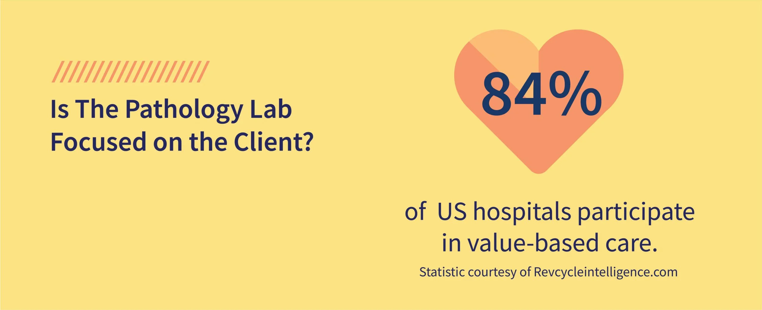
1. Are they focused on the client? 

With more healthcare systems moving to a value-based care model, patient-centered care is essential to ensure the seamless delivery of quick results and effective treatment plans while providing as much information as possible about your patient’s condition. It’s difficult to reassure your patient that their treatment is a priority when the turnaround time for lab results takes several weeks. 

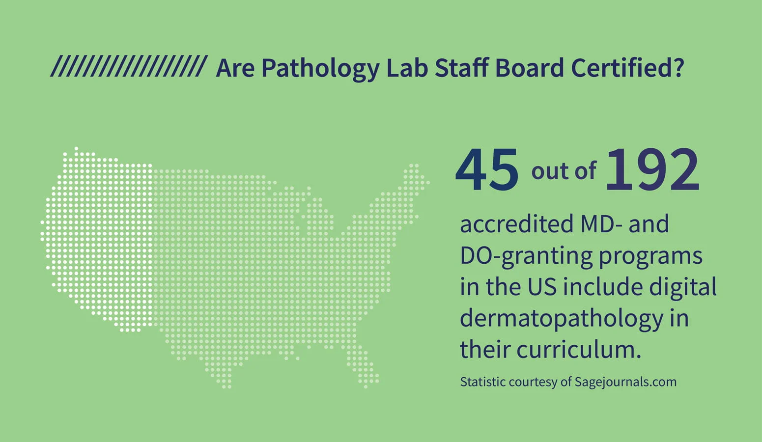
2. Are the lab techs board certified? 

Check to be sure that the lab technicians and specialists are educated and qualified to do their job. Although jobs as a general lab technician don’t require extensive college training, hands-on experience working in digital pathology is more specialized. 

As more labs implement digital technology into their lab processes, it’s important to clarify their expertise in operating the specialized equipment since it varies a great deal from handling glass slides and manually preparing lab test results.

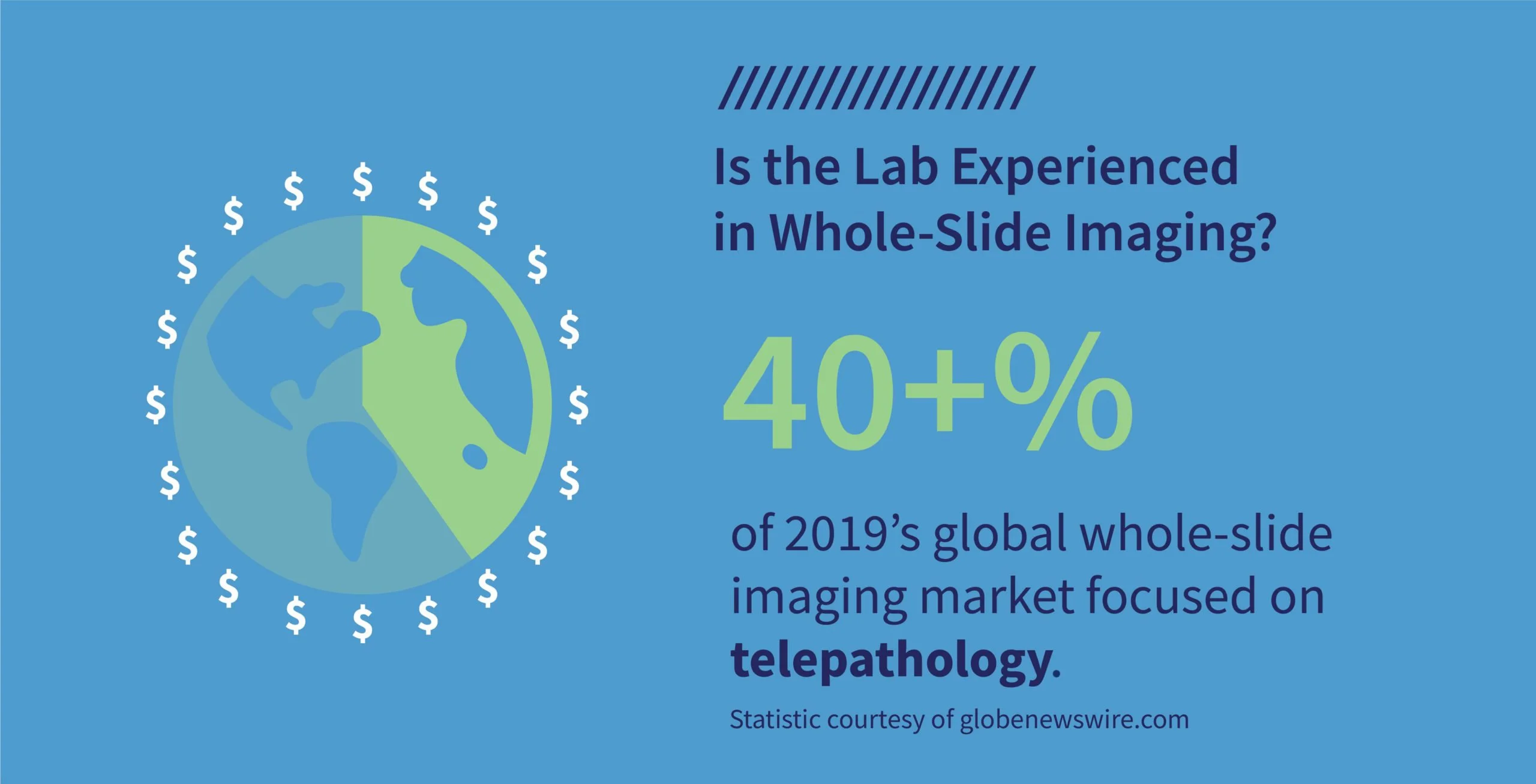
3. Do they have experience using whole-slide imaging?

Digital technology and whole-slide imaging is expanding throughout the dermatopathology industry, with many implementing digital innovations into their processes. As mentioned earlier, make sure the lab you rely on for fast and accurate results knows how to operate this new technology. 

Research conducted by Joann G. Elmore, MD, MPH, and her team concludes that navigating digital whole-slide imaging is different from traditional microscopy. In whole-slide imaging, the pathologist is not confined to a physical microscope that requires a glass slide viewed through a lens to view the tissue sample. 

“Rather, the digitized images of the histology tissue sections are viewed on a computer screen using a pointing device such as a mouse, trackpad, or dedicated console to manipulate location and magnification of the image (pan and zoom),” says the research team. “The technology may be easily adapted to virtual reality glasses. Given these important differences between digital WSI and traditional microscopy, adoption and effective use of WSI in clinical practice requires exposure to and training using this new format.”

4. What tests do they perform?

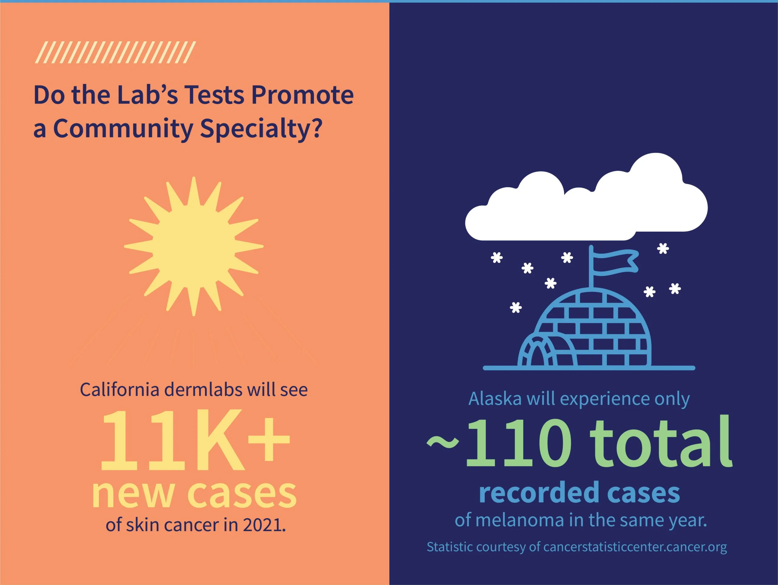
Based on the region, some labs have more experience performing certain tests that are more pronounced in your customer base than others. For example, a dermlab in California will likely see more tests for melanoma because it has the most cases of skin cancer (11,450 new cases this year) in the nation. By comparison, Alaska has 110 melanoma cases. 

If your practice specializes in treatments more common in the area, you want to partner with a lab that is proficient in those tests.

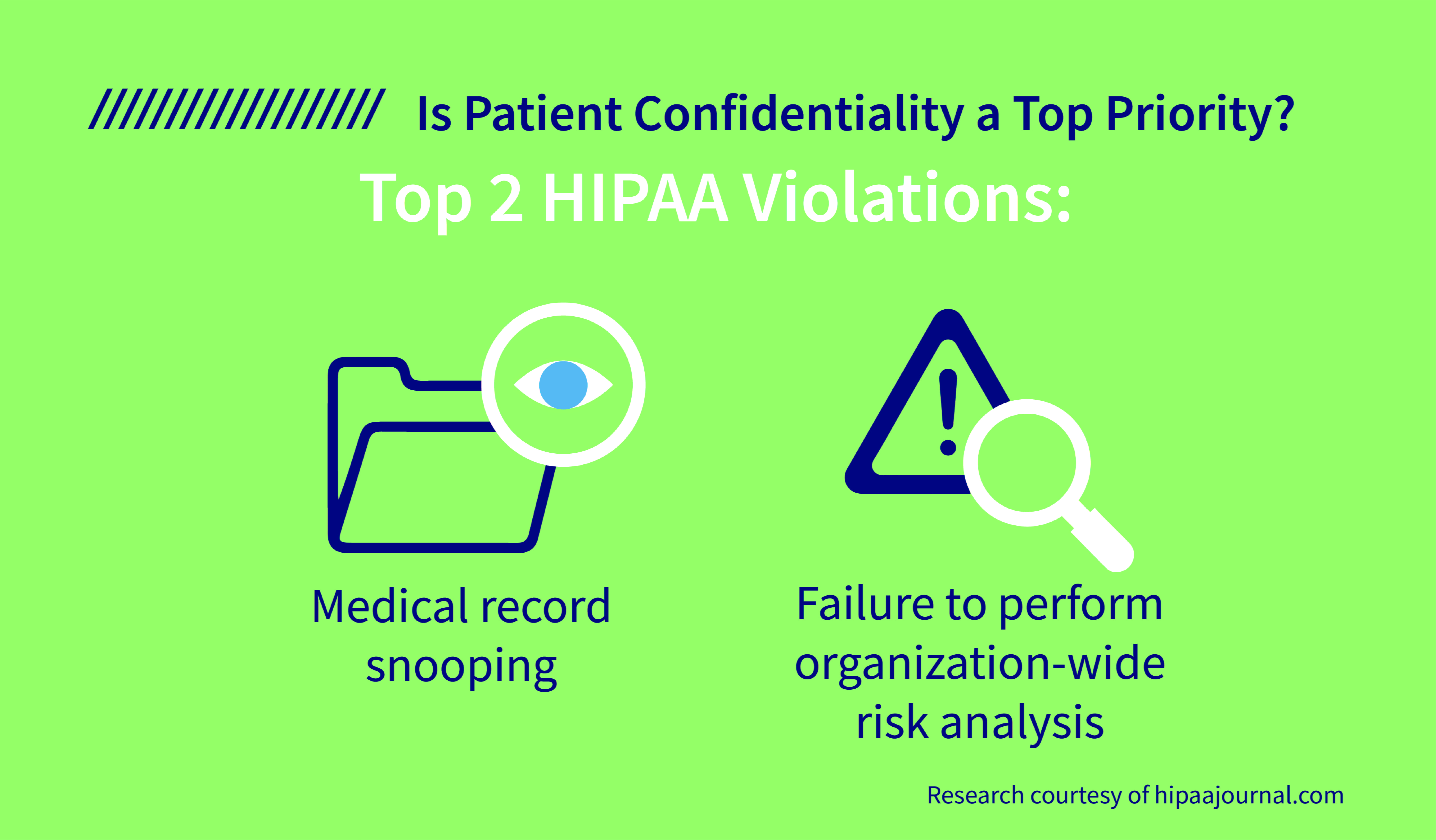
5. Is patient confidentiality a priority?

The wave of digital technology within the dermatopathology field has provided unprecedented access to patient information. With that abundance of data comes a higher risk of HIPAA violations. Be sure to ask how the lab processes and organizes testing samples and inquire about their procedures for sharing results. 

“Using an electronic health record or EHR system offers you much better control over information security,” says Stephen O’Connor. “What’s more, the electronic version of the patient’s chart is now more convenient to share with other concerned parties.” 

To expand your practice, you need a dermlab that offers immediate and secure access to digital slides, fully interpreted pathology reports uploaded directly into your patient’s EMR, and personalized service through your own dedicated dermpath team with the flexibility to collaborate with your existing dermatopathology provider. 

To learn more about what PathologyWatch digital dermatopathology lab services can do to help expand your practice, click here