Images shown are not intended to be used for the diagnosis or treatment of a disease or condition.
Sebaceous neoplasms are abnormal growths of cells originating in the oil glands of the skin. As part of the ongoing Digital Dermatopathology Digest series, Rajni Mandal, MD, a clinical research associate in dermatopathology for PathologyWatch, discusses the characteristics of sebaceous differentiation, up to and including sebaceous carcinoma.
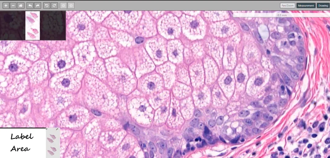
Sebaceous glands found in the dermis are formed by mature sebocytes and immature sebaceous cells. Mature sebocytes show evacuated cytoplasm, which can indent the nucleus in contrast to the immature cells that show increased nuclear-cytoplasmic ratio, sometimes with foamy cytoplasm.
“Neva sebaceous has an increased risk of basal cell carcinoma, trichoblastoma, and syringocystadenoma papilliferum,” says Rajni.
Sebaceous adenoma is defined as having greater than 50 percent mature sebaceous cells as compared to basaloid cells, in contrast to a sebaceoma. It can present as multiple nests in the dermis, with a predominance of the immature basaloid cells, as compared to the mature sebocytes.
The malignant counterpart of a sebaceous neoplasm is sebaceous carcinoma. It is most common in the eyelid, originating from the Meibomian gland. It can include the dermis as a proliferation of predominantly immature cells. In the epidermis, sebaceous cells—which are predominantly immature—can infiltrate in a pagetoid manner, which can mimic squamous cell carcinoma.
To learn more about sebaceous neoplasms, check the Digital Dermatopathology Digest video series, which provides detailed information and examples on a number of skin conditions, click here.