Teledermatology in the COVID Era

Despite the benefits touted by telemedicine enthusiasts, widespread adoption of this technology has lagged in the healthcare industry. However, with the onset of COVID-19, a new era of telemedicine has begun. Almost overnight, telemedicine became more accepted by regulators, providers, insurance companies, and patients. The combination of improved video technology, patient demand, and provider acceptance has eased restrictions and pushed telemedicine to the forefront in the medical industry. 

In addition, dermatopathology is following this trend. Regulations for the remote use of pathology have changed in light of COVID-19, making it possible for pathologists to work remotely and support the use of digital pathology technology for the duration of the pandemic.

With the universal adoption of video conferencing, 20 percent of all medical visits are happening via telemedicine. Here are a few reasons to consider teledermatology:

Convenience

Recent surveys show that a common reason over one-third of patients may prefer telemedicine visits is saving time by avoiding the commute to a provider’s office. For instance, telemedicine can reduce the amount of time away from work or the cost and headaches of arranging childcare. Patients can be productive while waiting for the video conference instead of spending time in the waiting room. Even older patients who might have struggled with technology adoption in the past are more likely to embrace telemedicine options to avoid the increased COVID complications and mortality associated with this at-risk population.

Accessibility

Dermatologists can now use traditional billing codes for teledermatology visits. Teledermatology is accessible even when providers or patients may be out sick but are not debilitated. Many people who test positive for COVID are asymptomatic and would still like to keep their appointments if possible. This helps control the inevitable cancellations required for patients with cold symptoms and need to rule out COVID or flu infection before presenting in your office.

Because of increased access provided by digital dermatopathology, companies like PathologyWatch can employ a national network of dermatopathology experts. Many areas do not have access to dermatopathologists and often rely on expensive locum tenens to provide specialty or overflow care. Also, it can take several weeks to get a second opinion on a case. With a digital workflow, digital slides can be more quickly accessed for consensus diagnosis or consultation.

Teledermatology also provides people living in rural areas or mobility challenges with easy access to medical care using synchronous live video-conferencing. The CDC indicates that telemedicine “can help reduce barriers to care for people who live far away from specialists or who have transportation or mobility issues.” As one dermatologist described, “Our specialty is a visual field, and there are many skin conditions which are diagnosable from looks alone.” Dermatology lends itself well to photographic consultations by primary care providers to dermatologists. 

Safety

During the COVID pandemic, the CDC established guidelines to keep healthcare workers and vulnerable patients safe during face-to-face visits. Despite these precautions, healthcare workers are still seven times more likely to develop severe COVID-19 infection than individuals performing nonessential jobs.

Teledermatology encourages a workflow that keeps providers and their staff safe from contracting diseases from patients or other staff members. Also, video conferencing can allow for the outsourcing of specific tasks, such as a scribe. This can help dermatology clinics by having a safe pool of healthcare workers that don’t have a higher risk of developing COVID-19. 

Patient Satisfaction

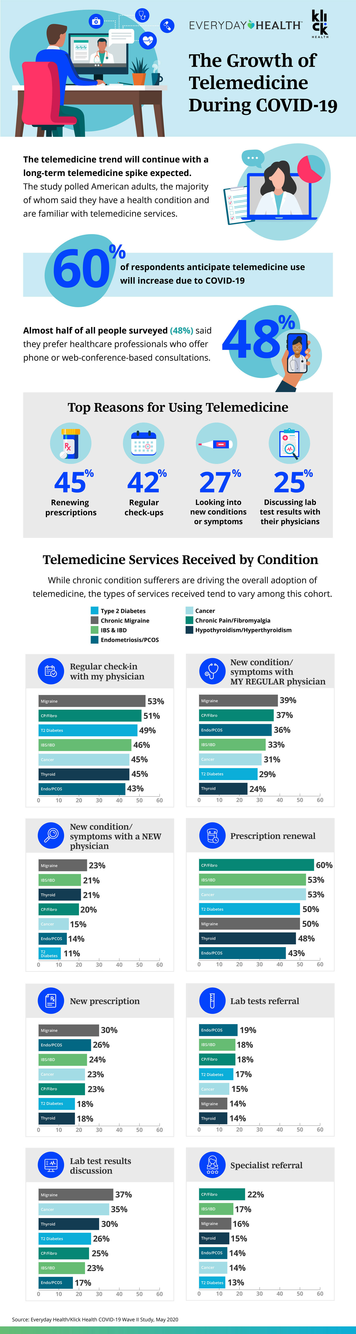
Up to 48 percent of people prefer providers who offer telemedicine visits. A consumer survey revealed that 75 percent of patients who participated in virtual care were very or completely satisfied with the experience. 

Meanwhile, telemedicine can level the socioeconomic playing field. Access Derm is an AAD-supported teledermatology program where volunteer dermatologists can help underserved populations who don’t have access to a dermatologist. It helps provide critical preventive care and second opinions, improving the quality of life and reducing the expense of future treatments by diagnosing skin cancer at earlier stages, for example. 

When to Consider Teledermatology

Consider teledermatology options for follow-up visits that focus primarily on counseling rather than diagnosis. With these types of patients, rapport has already been established through in-person visits, and with straightforward diagnoses—like acne, atopic dermatitis, and psoriasis—much of the return visit focuses on review of lab work, medication counseling, and treatment changes if patients are not responding to first-line therapies. In particular, iPLEDGE patients or their caregivers, who are required to have monthly visits for the duration of therapy, may save significant time through a teledermatology visit. 

While not all visits are possible through video conferencing, making healthcare convenient can encourage more people to seek regular consultations and take better care of themselves.

Tips to Open Your Own Dermatology Practice

You’ve done it. You’ve decided to take the plunge and open your own dermatology practice. Congratulations! But take care: With about 20 percent of new small businesses failing in the first year, it’s key to learn from those who have traveled the road to health services self-employment before you. So before you shop for locations and window treatments, here are five strategic tips to ensure your open-for-business experience is healthy and successful. 

Secure your capital. 

It’s true: For a new business to succeed, it really does take money to make money. Experts estimate that almost 30 percent of new businesses fail because they run out of cash. In fact, results collected by Investopedia show that two of the four most common reasons why small businesses fail involve cash-flow problems—mainly insufficient budgeting and a lack of sufficient capital. 

For Daniel Lambert, CEO of PathologyWatch, exploring various forms of capital was pivotal to startup success. “Look at raising from different asset classes: private equity, growth equity, sovereign wealth funds, PE firms, family offices and cash-rich investors. There are many sources of capital that are often overlooked,” he advises. 

Once you’ve formalized your vision and bolstered it with a satisfactory cash flow, it’s essential to hire key players who share your vision and offer expertise in fundamental skills. 

Hire people with office management, business, and legal expertise.

You may be an expert at differentiating herpes zoster from acne vulgaris, but do you know the latest HIPAA compliance requirements or how to handle payroll tax? Your staff will need both clinical and nonclinical support, so one of your first steps should be hiring those with hands-on management, legal structure, and compliance experience. 

“Choosing the right legal structure for your medical practice is one of the most important business decisions you’ll make,” says Jack Wolstenholmhead of content marketing at LeverageRx. “Deciding whether to establish a sole proprietorship or a more complex legal structure must be thought through wisely. It requires calculating both the costs and benefits to your practice, and factoring in your risk tolerance to liability.”

Staying ahead of the game in one of the most highly regulated industries could mean the difference between staying open or becoming a statistic. 

Establish your specialized services.

What is your specialty? For instance, if you’re setting up a practice in an area with a year-round sunny climate, specializing in skin-cancer treatment could be your focus. If you are unsure, check out practices in the area. Remember that while you are providing healthcare, you are also a small business owner. That means keeping an eye on your competitors. To narrow down prospective areas, call some practices to check on waiting times for an appointment for new patients. If the wait time is longer than two weeks, there’s plenty of room for another doctor. 

“Public demographic information can tell you about future growth in different communities,” say experts at The Dermatologist. “If you expect to attract patients from many different communities, look at traffic flow to make sure your practice is easily accessible.” 

Your expertise will help build your patient base, but it’s the mindset of a small business owner that will help keep your doors open for business.

Start by investing only in the essential equipment and space.

With this new cash flow, it’s tempting to overspend during the initial stages of business development. Before you schedule the interior decorator, look at your financial overhead with a long-term perspective. This endeavor is going to put your ability to maximize cash flow to the ultimate test. 

One area where you can minimize overhead expenses is usable physical space. Experts recommend starting small and leaving room for expansion. For example, when Jerome Obed, DO, a board-certified dermatologist who runs Broward Dermatology and Cosmetic Specialists in Fort Lauderdale, Florida, opened his new practice, he only prepared two out of his four exam rooms. When his patient numbers increased, he furnished the remaining two exam rooms in his office. 

The process of gradually building up at the same rate of acquiring new patients applies to investing in equipment as well. When you’ve determined your specialty, those are the pieces that should be purchased first. To control startup costs, we’ll discuss later the options of outsourcing certain functions to free up funds to purchase essential equipment.

Along with medical equipment, your IT system is an essential part of your office operation—and one of the most expensive. “You’ll need a practice management system for demographic information, scheduling, and billing, and an electronic health record (EHR) system for patient information,” say the staff at The Dermatologist.

Consider outsourcing functions.

Using a third-party service for billing or payroll tasks is a common practice for medical offices of all sizes. Services like PathologyWatch can manage both the business and technology aspects of digital pathology technology. That means your startup can offer all of the services you need to grow your new practice without having to invest in your own laboratory equipment and lab staff. 

You may not know what lies ahead when you open your own dermatology practice. But by talking with people who share in your journey for a private dermatology practice and following these five proven tips, you can learn ways to minimize risk while maximizing opportunities for growth.

How Well Do You Know Basal Cell Carcinoma?

Images shown are not intended to be used for the diagnosis or treatment of a disease or condition.

In this episode of Digital Dermatopathology Digest, Rajni Mandal, MD, a dermatopathologist at PathologyWatch, discusses the most common dermatological malignancy—basal cell carcinoma (BCC)—including how common it is, its warning signs, and characteristics of its various forms.


How Common Is BCC?

Prolonged exposure to UV sunlight or indoor tanning contributes to this and other forms of skin cancer, which triggers mutations in p53, PTCH (sonic hedgehog pathway), and SMO gene. Research shows that four million Americans are diagnosed with basal cell carcinoma every year. Most cases occur in people over the age of 50, and men are more likely to develop BCC than women. 

What Are The Most Common Subtypes?

“There are two common types: nodular and superficial,” explains Rajni. She also points out the aggressive types of basal cell carcinoma include infiltrative, micronodular (which presents characteristics similar to those of the nodular subtype, except it’s comprised of multiple nodules), morpheaform, and basosquamous subtypes.

What Are Differences between Superficial and Nodular BCC?

Features of superficial BCC include clusters of basaloid cells descending from the epidermis with no dermal invasion.

The most common subtype, nodular basal cell carcinoma has islands of cells with peripheral palisading and a haphazard arrangement of the more central cells. 

What Are the Characteristics of Morpheaform BCC?

Also known as sclerosing basal cell carcinoma, this aggressive subtype has spiky, basaloid, thin cell strands that chase down the dermis with its dense fibrous stroma. Differential diagnoses may include metastatic cancer, microcystic adnexal carcinoma, and desmoplastic trichoepithelioma. 

What Are Clinical Symptoms of BCC?

Patients with basal cell carcinoma may experience:

  1. Sores that don’t heal
  2. Patches of skin that remain irritated and red
  3. A bump of skin that may resemble a mole but appears shiny,  pink, or pearl-like in appearance
  4. An area where skin looks shiny and taut with poorly defined borders and a scar-like appearance

These symptoms are most likely to occur in areas often exposed to the sun, such as one’s face, neck, scalp, back, shoulders, ears, and chest. Other risk factors include Gorlin syndrome and nevus sebaceus.

Whether you’re in residency, studying for board exams, or a practicing dermatologist looking to stay sharp, the Digital Dermatopathology Digest video series is your informational and convenient source for dermatopathology review. Find the full series here.

Improving Communication and Clinicopathological Correlation with the Dermpath Lab

Clinicopathological correlation is the thought process that combines both gross and microscopic information to determine the most probable diagnosis. Being captivated by seeing the clinical image and pathology slide side by side is a common sensation for physicians feeling drawn to dermatology. This marriage of clinical and pathologic findings is also fundamental to resident and continuing medical education for dermatologists.

Clinicopathological correlation helps to confirm clinical suspicions and to provide more information in more obscure clinical cases. A nine-year study of nearly 4,000 skin biopsy specimens reported that 23.2 percent of the pathological diagnoses were inconsistent with the clinical diagnoses, suggesting room for diagnostic accuracy improvement. 

Studies suggest that improved clinical and pathologic correlation can help bridge that gap. By reviewing your own biopsy slides, providing accurate clinical information to your pathologist, and correlating together on challenging cases, your dermatology practice can continue to give patients the quality care they deserve.

Back to Your Roots: Reviewing Biopsy Slides

While only one out of five dermatologists reportedly read their own slides, most dermatologists prefer to review their slides. Dermatologists also receive significant training in dermatopathology during residency, more than twice that of their general pathology colleagues, and dermatology journals include more articles on dermatopathology. While avoiding the liability of reading their slides, many dermatologists enjoy keeping up their skills, confirming their clinical findings, or obtaining additional information needed to secure a diagnosis.

However, the traditional dermatopathology workflow may prevent dermatologists from reading or reviewing their own slides. Less than 25 percent of dermatologists have an in-house lab; if sending to an outside lab, it can take up to two weeks to turn around slides and even results. By using a lab that utilizes a digital pathology workflow, like PathologyWatch, dermatologists have quicker and easier access to review their own pathology slides or those of their colleagues, providing helpful information when planning surgical excisions or Mohs procedures, for example. 

Don’t Rule Out the Dermatopathology Requisition

Research studies emphasize the importance of clinical information in making accurate pathologic diagnoses, particularly in dermatopathology. In a recent Dialogues in Dermatology podcast, an important study was reviewed, indicating that dermatopathologic diagnostic accuracy is 53 percent when no clinical information is provided; accuracy improves to 78 percent when information is provided. Another study emphasized the importance of continued correlation in difficult cases, noting that repeat biopsy with additional CPC improved the concordance of clinical and pathologic diagnoses further. 

Providing dermoscopic images and essential diagnostic criteria for melanoma—like size, partial or complete biopsy, and evolution of a lesion—can also influence the pathologic diagnosis, likely resulting in improved patient outcomes. Dermatologists are particularly well-trained in providing helpful information on their requisition forms.

Tips on communicating effectively with the lab include having clinicians (rather than medical assistants) complete (or dictate) findings on the requisition, providing a helpful list of differential diagnoses, and giving other relevant clinical information and photographs. 

The Power of a Simple Phone Call

Lastly, in challenging cases, or simply where vital information was left out, a quick text or phone call with an experienced dermatopathologist can be invaluable in clinching the diagnosis. 

Sometimes you may find that important clinical information, such as a genetic disease or patient age, is left off the requisition, potentially skewing a pathologic diagnosis. This critical information needs to be passed on to the consultant, just as key clinical information from a patient may help in assuring your clinical diagnosis. 

If a dermatologist reads their own slides, consulting with a network of expert dermatopathologists like PathologyWatch on difficult cases can improve diagnostic accuracy.

While physicians are often pressed for time in the clinic, taking time to review pathology slides, providing accurate information to your dermatopathology lab, and interacting with consultants directly to make important clinicopathological correlations will ultimately result in time savings and better patient outcomes by making an accurate diagnosis. 

Can You Identify an Epidermal Inclusion Cyst?

Images shown are not intended to be used for the diagnosis or treatment of a disease or condition.

An epidermal inclusion cyst is the most common cutaneous cyst, often called a sebaceous cyst, which is actually a misnomer. The center of epidermoid cysts almost always contains keratin and not sebum. This keratin often has a “cheesy” appearance. They also do not originate from sebaceous glands; therefore, epidermal inclusion cysts are not truly sebaceous cysts.

In this episode of Digital Dermatopathology Digest, Rajni Mandal, MD, dermatopathologist at PathologyWatch, explains the common characteristics of an epidermal inclusion cyst.

“An epidermal inclusion cyst has an epidermis-like lining with a granular layer,” explains Mandal—as opposed to a nongranular layer, seen in a pilar cyst or steatocystoma. “Sometimes there is a connection with the overlying epidermis known as a punctum.” 

Mandal goes on to mention features, the lack of which can help distinguish epidermal inclusion cysts from other common cysts, including dermoid, pilar, and vellus cysts.

Ruling Out a Dermoid Cyst

A dermoid cyst has cell walls that contain hair follicles, sweat glands, and other multiple adnexal skin structures. By contrast, an epidermal inclusion cyst lacks adnexal structures.

Ruling Out a Pilar Cyst

What makes a pilar cyst unique is that it arises from the epithelium between the sebaceous gland and the arrector pili muscle. They are lined by stratified squamous epithelium without a granular cell layer, similar to what is seen in the outer root sheath of the hair follicle, and filled with keratin.

Often presenting themselves as a round, dome-like bump, a pilar cyst is typically firm to the touch but not painful for the patient, while an epidermal inclusion cyst may become inflamed and painful to the touch. 

Ruling Out a Vellus Hair Cyst

If small hairs appear in the vellum, with small red or brown bumps and a smooth dome shape, and often occur over the sternum, it is likely a vellus hair cyst.

Whether you’re in residency, studying for board exams, or a practicing dermatologist looking to stay sharp, the Digital Dermatopathology Digest video series is your informational and convenient source for dermatopathology review. Find the full series here.