Images shown are not intended to be used for the diagnosis or treatment of a disease or condition.
In order to diagnose pathologic skin conditions, it’s important to first be able to recognize what constitutes normal skin and variants. In this Digital Dermatopathology Digest episode, Rajni Mandal, MD, uses a digital pathology image viewer to offer a quick summary of key characteristics in normal skin. Here are some video highlights.
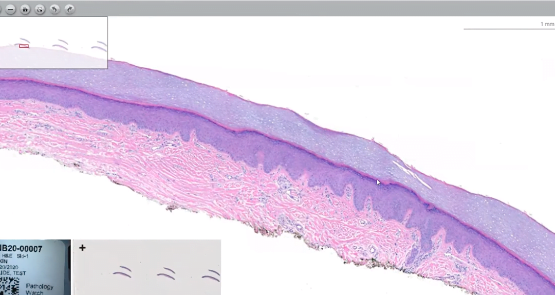
Skin Anatomy
The body’s largest organ takes on a variety of appearances depending on the anatomic location. The two outermost layers—the epidermis and the dermis—work closely together. The epidermis, or outer layer, typically has four or five distinct layers of cells but lacks blood vessels or nerve endings.
The inner layer, or the dermis, consists of pink collagen fibers, which are connective tissue that supports, nourishes, and strengthens the epidermis. Also contained within the dermis are key structures such as nerve endings, blood vessels, lymph glands, hair follicles, and sweat glands.
“The dermis can vary in thickness based on location,” says Mandal. Skin of the back and trunk shows more compact collagen fibers and a thicker dermis compared to facial skin. Acral skin shows compact hyperkeratosis of the stratum corneum. Solar elastosis, seen in areas of sun damage, has a gray-purple hazy color and is a collection of excess elastic fibers.
Distinguishing Features of Adnexal Structures
Sebaceous glands have foamy cytoplasm and often attach directly to the hair follicle.
Deeper in the dermis, you can find eccrine sweat glands, which occur over most of your body and open directly onto the surface of your skin. “Eccrine ducts have a two-layer epithelium versus the eccrine glands, which have a single layer epithelium,” explains Mandal. Adipocytes are also found admixed around the glands.
By contrast, apocrine sweat glands are found in the axilla, nipple, ear canal, eyelid, scalp, and genital areas. Very round nuclei, eosinophilic cytoplasm, and eosinophilic secretions within the lumen are clues to apocrine glands.
If you enjoyed this summary of identifying normal skin and variants, or if you are preparing for boards and want to test your dermatopathology skills, check out this and other Digital Dermatopathology Digest instructional videos here.