How to Explain Dermatopathology to Your Patients

According to IBISWorld, there are an estimated 5,391 dermatology businesses in the United States, serving millions of individuals who understand the importance of taking care of their skin. But when it comes to the science behind diagnosing and treating skin conditions, some patients may feel lost, confused, or scared.

Explaining what dermatopathologists do can entail a lot of information for a patient, so let’s examine some simple ways to discuss what dermatopathology is, including education, training, and specialties, as well as the advancement of digital dermatopathology and how it is changing the industry.

What Is Dermatopathology?

Dermatopathology is a specialty in the field of dermatology. The term itself is a combination of dermatology and pathology, meaning the study of both the skin and diseases thereof. Pathologists work in every field of medicine, providing insight on diseases and patient care—in the field of dermatology, this is the responsibility of the dermatopathologist—or “dermpath,” as they are sometimes known in the industry.

All dermatopathologists hold a medical degree in dermatology or pathology, with a subspeciality in dermatopathology. In order to become board certified, doctors must take an exam in both their specialty and subspecialty, as well as participate in developmental exercises in order to retain that certification.

Dermatopathology Services

Rather than interacting directly with patients, dermatopathologists analyze tissue samples that are sent over by doctor’s offices to provide additional information. A nine-year study by the National Library of Medicine found an increasing trend in the complexity of dermatopathology cases after reviewing 8,173 cases at a tertiary care academic center. 

Dermpaths are able to examine not only skin samples but also hair and nail tissue samples, after which they generate a report based on their findings. This is typically done when a dermatologist or primary care physician wants to confirm an initial diagnosis made during a physical examination of the potentially affected area, and requests to have a sample tested by a dermatopathologist for more information.

In studying these samples, dermatopathologists are able to better identify a wide variety of skin conditions, including skin cancer, eczema, and psoriasis. Examining samples under a microscope allows dermatopathologists to see them on a cellular level, providing a closer perspective of a patient’s potential diagnosis.

Traditionally, dermatopathology was done using microscopes, but technological advances have led to the increasing use of digital dermatopathology, in which samples are processed and digitized, providing doctors with more readily available access to a patient’s slides. This process minimizes the time it takes to send a sample, receive the results, and make a diagnosis based on those results, which can be life-saving in more severe cases.

Uses and Benefits of Digital Dermatopathology

Digital dermatopathology helps medical professionals to continue providing patients with the level of care they require while offering clear advantages for your dermatology clinic. These include access to quality clinical care for rural markets, shorter diagnostic times than traditional microscopy, and equivalence to glass slides in terms of quality, with a major discordance rate of only 0.4 percent between whole-slide imaging and microscopy.

Digital dermatopathology can also reduce the time it takes between receiving a slide and generating a report. In some cases, hospitals staff saw an average turnaround time reduced from two weeks to two days. Elsewhere, the streamlined digital process saves the staff an hour a day on manual tasks.

Now that you have some ideas of how to explain what dermatopathology is to your patients, contact the experts at PathologyWatch to help with all your digital dermatopathology needs.

Is Your Dermlab a Good Fit? 5 Questions to Ask a Dermatopathology Lab

5 questions to ask your pathology lab
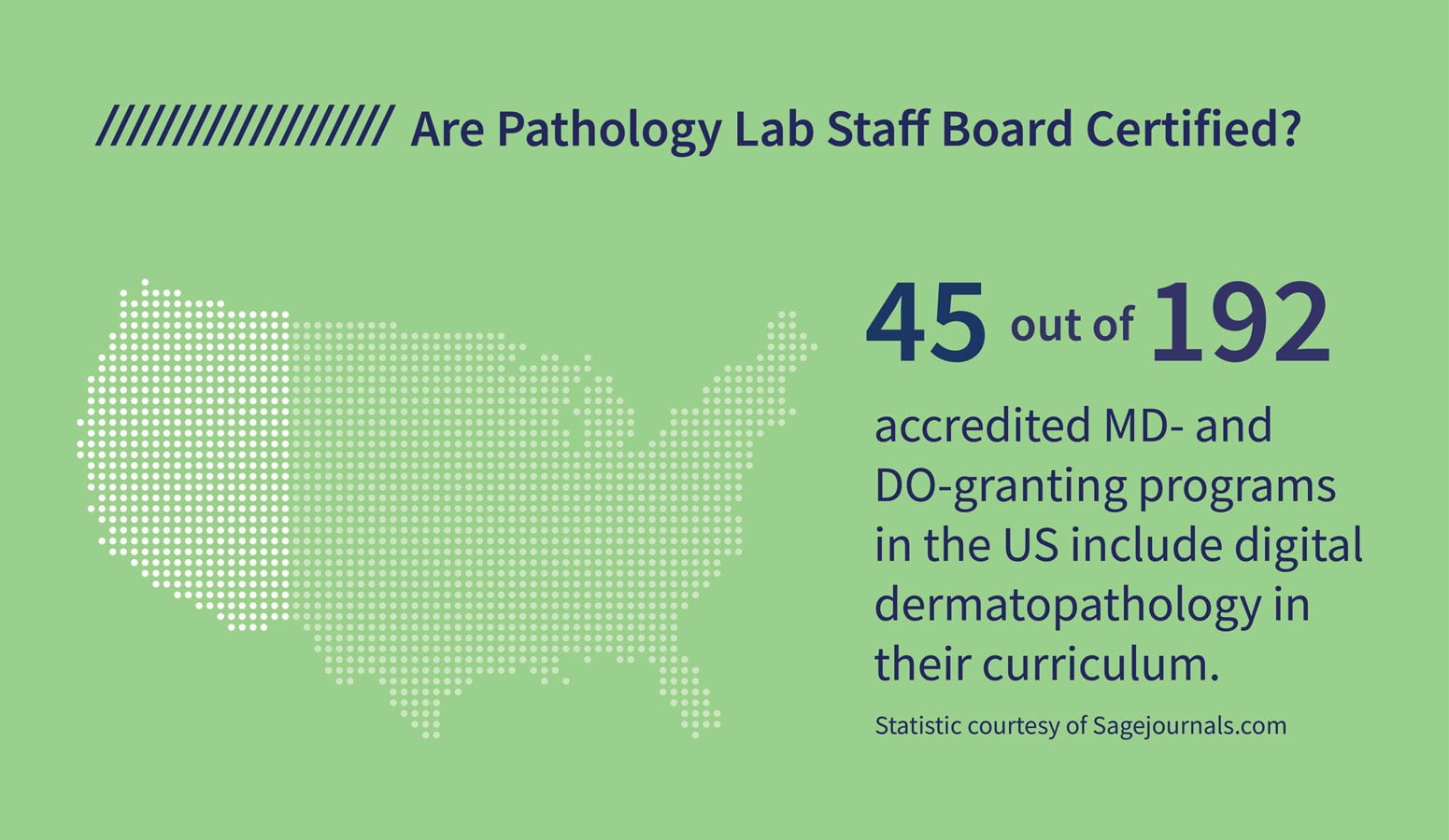
are staff board certified
focused on the client
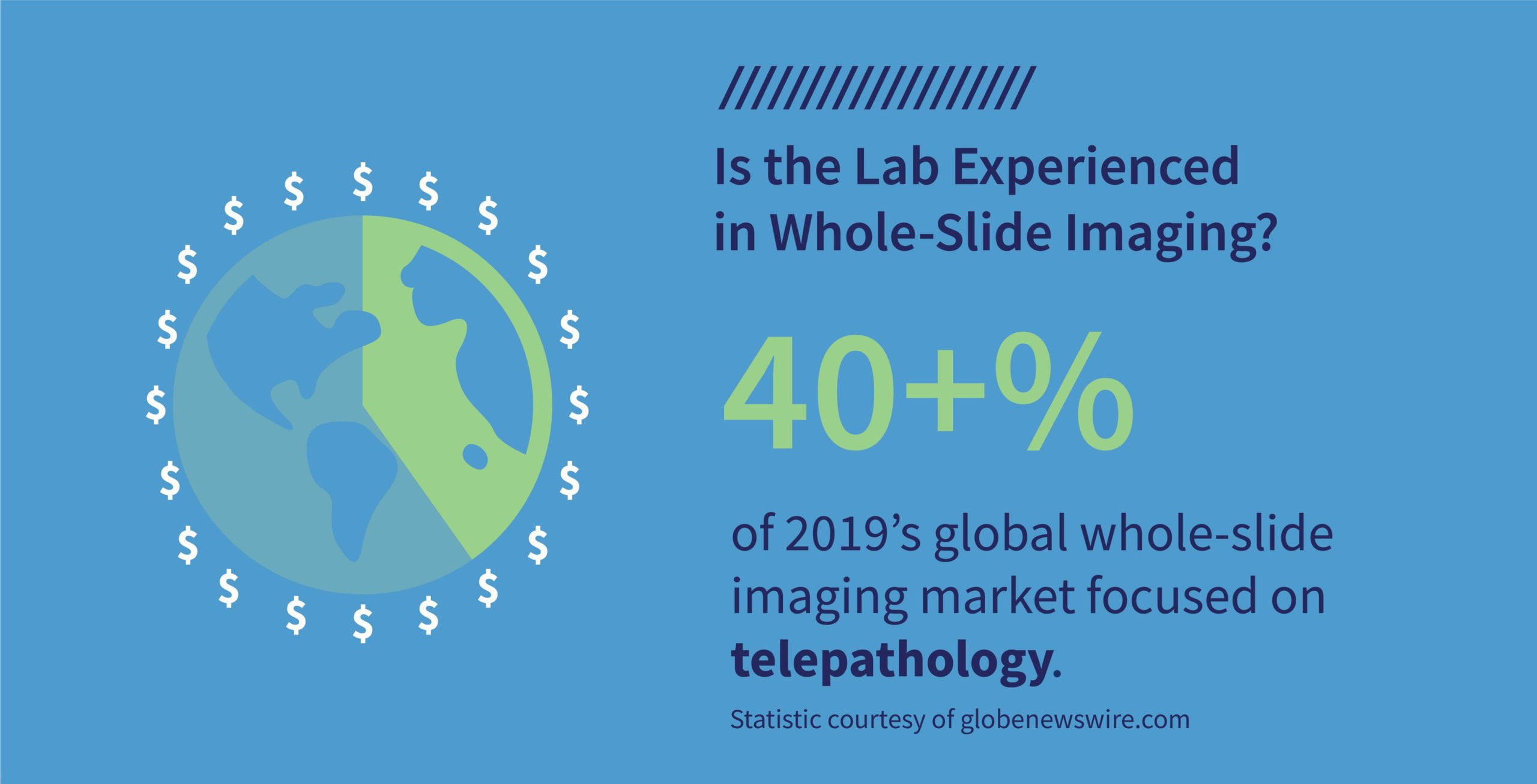
whole-slide image experience
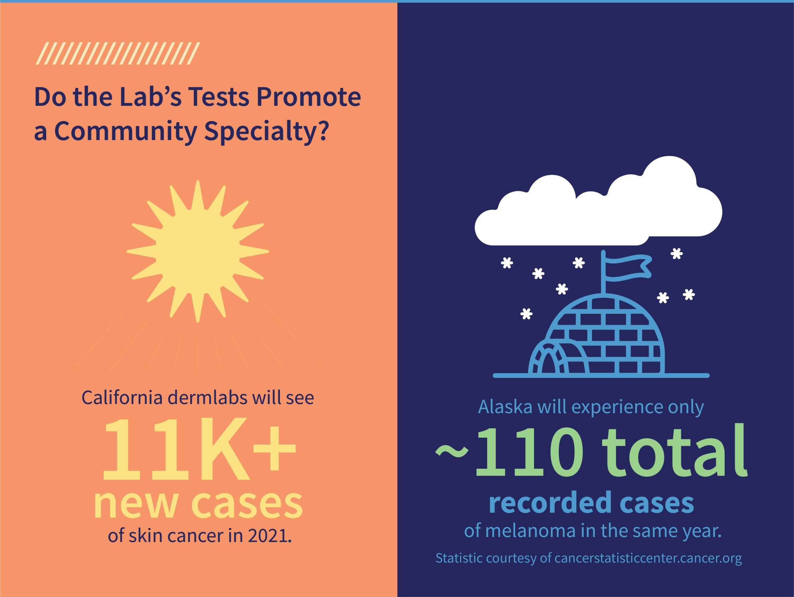
promote community specialty
prioritize confidentiality
contact pathologywatch today
Are you searching for a new dermatopathology lab? With the onset of digital pathology, finding a lab that is not only equipped with this innovation but also qualified to process whole-slide images swiftly yet accurately may take some time. 

An NIH study found that three out of four respondents agreed that accurate diagnoses can be made with this technology, and over half (59 percent) agreed that the benefits of whole-slide imaging outweigh any concerns. If you plan to expand your practice to incorporate whole-slide imaging into your process, finding the right dermlab is an important first step. 

To ensure the ideal match, here are five questions to ask dermatopathology labs. 

1. Are they focused on the client? 

With more healthcare systems moving to a value-based care model, patient-centered care is essential to ensure the seamless delivery of quick results and effective treatment plans while providing as much information as possible about your patient’s condition. It’s difficult to reassure your patient that their treatment is a priority when the turnaround time for lab results takes several weeks. 

2. Are the lab techs board certified? 

Check to be sure that the lab technicians and specialists are educated and qualified to do their job. Although jobs as a general lab technician don’t require extensive college training, hands-on experience working in digital pathology is more specialized. 

As more labs implement digital technology into their lab processes, it’s important to clarify their expertise in operating the specialized equipment since it varies a great deal from handling glass slides and manually preparing lab test results.

3. Do they have experience using whole-slide imaging?

Digital technology and whole-slide imaging is expanding throughout the dermatopathology industry, with many implementing digital innovations into their processes. As mentioned earlier, make sure the lab you rely on for fast and accurate results knows how to operate this new technology. 

Research conducted by Joann G. Elmore, MD, MPH, and her team concludes that navigating digital whole-slide imaging is different from traditional microscopy. In whole-slide imaging, the pathologist is not confined to a physical microscope that requires a glass slide viewed through a lens to view the tissue sample. 

“Rather, the digitized images of the histology tissue sections are viewed on a computer screen using a pointing device such as a mouse, trackpad, or dedicated console to manipulate location and magnification of the image (pan and zoom),” says the research team. “The technology may be easily adapted to virtual reality glasses. Given these important differences between digital WSI and traditional microscopy, adoption and effective use of WSI in clinical practice requires exposure to and training using this new format.”

4. What tests do they perform?

Based on the region, some labs have more experience performing certain tests that are more pronounced in your customer base than others. For example, a dermlab in California will likely see more tests for melanoma because it has the most cases of skin cancer (11,450 new cases this year) in the nation. By comparison, Alaska has 110 melanoma cases. 

If your practice specializes in treatments more common in the area, you want to partner with a lab that is proficient in those tests.

5. Is patient confidentiality a priority?

The wave of digital technology within the dermatopathology field has provided unprecedented access to patient information. With that abundance of data comes a higher risk of HIPAA violations. Be sure to ask how the lab processes and organizes testing samples and inquire about their procedures for sharing results. 

“Using an electronic health record or EHR system offers you much better control over information security,” says Stephen O’Connor. “What’s more, the electronic version of the patient’s chart is now more convenient to share with other concerned parties.” 

To expand your practice, you need a dermlab that offers immediate and secure access to digital slides, fully interpreted pathology reports uploaded directly into your patient’s EMR, and personalized service through your own dedicated dermpath team with the flexibility to collaborate with your existing dermatopathology provider. 

To learn more about what PathologyWatch digital dermatopathology lab services can do to help expand your practice, click here

Digital Dermatopathology Digest: Identifying Hair Follicle Tumors

Images shown are not intended to be used for the diagnosis or treatment of a disease or condition.

There are a number of different hair follicle tumors, each with its own distinguishing features. In the Digital Dermatopathology Digest series, Rajni Mandal, MD, clinical research associate in dermatopathology for PathologyWatch, discusses these different variants in greater detail.

Trichilemmal cells come from the outer root sheath of the hair follicle. The cells show abrupt keratinization and pink to clear cytoplasm with a lack of keratohyalin granules.

Trichilemmoma cells have a lobular proliferation of cells that can push into the dermis. They can also present as pink to clear due to glycogen, and there can be peripheral palisading at the periphery of the islands as well as a thick pink basement membrane deposition. Multiple trichilemmomas are associated with Cowden syndrome.

Trichoadenoma cells can have multiple epithelial islands of keratin-filled cysts in the dermis. These cysts can frequently rupture, shoring a foreign body giant cell reaction to keratin fibers. Cyst islands are lined by epithelium, and the majority of the squamous epithelium lacks keratohyalin granules.

Trichoblastoma cells typically have multiple blue islands with fibrotic stroma and are associated with nevus sebaceous.

Cells on the trichofolliculoma, fibrofolliculoma, and trichodiscoma spectrum are all different stages of the same tumor. They can present with branches of anastomosing epithelium originating out of the hair follicle and often occur in small antigen hair follicles.

  • In a fibrofolliculoma, there can be a proliferation of fibrous cells or fibrous stroma, which can have a concentric pattern around the tumor.
  • Fibrous proliferations are most prominent in a trichodiscoma.
  • Cells can show peripheral palisading, but there is no retraction artifact or myxoid stroma surrounding them, which would be seen in basal cell carcinoma.
  • Multiple fibrofolliculomas and trichodiscomas are commonly associated with Birt-Hogg-Dubé syndrome.

The Digital Dermatopathology Digest series provides educational information for both students and professionals on a number of topics. To view the complete series, click here.

Digital Dermatopathology Digest: Identifying Sebaceous Neoplasms 

Images shown are not intended to be used for the diagnosis or treatment of a disease or condition.

Sebaceous neoplasms are abnormal growths of cells originating in the oil glands of the skin. As part of the ongoing Digital Dermatopathology Digest series, Rajni Mandal, MD, a clinical research associate in dermatopathology for PathologyWatch, discusses the characteristics of sebaceous differentiation, up to and including sebaceous carcinoma.

Sebaceous glands found in the dermis are formed by mature sebocytes and immature sebaceous cells. Mature sebocytes show evacuated cytoplasm, which can indent the nucleus in contrast to the immature cells that show increased nuclear-cytoplasmic ratio, sometimes with foamy cytoplasm.

“Neva sebaceous has an increased risk of basal cell carcinoma, trichoblastoma, and syringocystadenoma papilliferum,” says Rajni.

Sebaceous adenoma is defined as having greater than 50 percent mature sebaceous cells as compared to basaloid cells, in contrast to a sebaceoma. It can present as multiple nests in the dermis, with a predominance of the immature basaloid cells, as compared to the mature sebocytes.

The malignant counterpart of a sebaceous neoplasm is sebaceous carcinoma. It is most common in the eyelid, originating from the Meibomian gland. It can include the dermis as a proliferation of predominantly immature cells. In the epidermis, sebaceous cells—which are predominantly immature—can infiltrate in a pagetoid manner, which can mimic squamous cell carcinoma.

To learn more about sebaceous neoplasms, check the Digital Dermatopathology Digest video series, which provides detailed information and examples on a number of skin conditions, click here.

Digital Dermatology Digest: Identifying Syringomas

Images shown are not intended to be used for the diagnosis or treatment of a disease or condition.

If you happen across a small cluster of brown, yellow, or pink bumps (or papules), around the eyes or on the neck, it may be a case of syringomas. In this episode of Digital Dermatopathology Digest, Rajni Mandal, MD, clinical research associate in dermatopathology for PathologyWatch, briefly discusses the distinct characteristics of this benign growth of the sweat gland. 

According to Rajni, syringomas are common in the periorbital and genital regions. Growths can be associated with Down’s syndrome, and disseminated, solitary, or eruptive types sometimes indicate Nicolau-Balus syndrome. 

“Note that the eccrine ducts have an eccentric lumen and can have a tadpole shape,” says Rajni, referring to a common variant of syringomas. “And a clear cell syringoma is often associated with diabetes.” 

Another type of syringomas, a mixed tumor called chondroid syringoma, is common in elderly males and appears on the head and neck. It’s characterized by a circumscribed dermal tumor, and it also appears as an apocrine/eccrine proliferation in fibrous, myxoid, or chondroid stroma. 

“Women, especially of Japanese heritage, are affected more than men,” explains the American Osteopathic College of Dermatology. “Syringomas tend to first erupt in adolescence, but could start at any age.” The AOCD adds that young people may be predisposed to “eruptive syringomas,” wherein the growths occur suddenly.  

Whether you’re in residency, studying for board exams, or a practicing dermatologist looking to stay sharp, the Digital Dermatopathology Digest video series is your informational and convenient source for dermatopathology review. Find the full series here.

PathologyWatch Ranked Top MedTech Innovator of the Year

PathologyWatch was recently ranked by MedTech Innovator as one of the top 50 most transformative device, diagnostic, and digital health technologies from around the globe. PathologyWatch will participate in a flagship, four-month Showcase and Accelerator program that offers unparalleled visibility and access to some of the medtech industry’s leading manufacturers, providers, investors, and other industry stakeholders. 

“Our goal at MedTech Innovator is to find the most promising medical innovations and make sure they actually reach the patients who need them,” said Paul Grand, CEO of MedTech Innovator. “We are thrilled with the caliber of the startups participating in this year’s cohort and we look forward to providing them with the resources and mentorship they need to succeed.”

Since 2017, PathologyWatch has been an innovative leader in intuitive, easy-to-implement digital dermatopathology solutions that aim to streamline the diagnostic process while improving patient care models. 

Compared to its competitors, PathologyWatch focuses on enhancing services available in outpatient clinics by offering a fully interfaced EMR reporting process, access to top dermatopathologists with the option to interpret and bill for their cases, and provide their clients 24/7 access to digital slides. 

“I believe PathologyWatch is the future of dermatopathology for dermatologists. The group combines academic expertise with the efficiency of a private lab. It is a huge timesaver for me and my staff,” said April Larson, a certified dermatologist, and VP of client experience and advisory board at PathologyWatch. “By reviewing my own cases, I am able to make better clinical decisions for my patients. It’s a win-win for everybody.”

With PathologyWatch, clinics can significantly reduce the time it takes from completing the patient examination to diagnosis with high-quality reads that lead to improved patient-provider relationships in shared medical decisions. What typically takes two weeks to receive a diagnosis can now potentially take two days through the efficient workflow that PathologyWatch has developed.

The 50 companies selected by MedTech Innovator will compete in the final competition at The MedTech Conference in September 2021. 

To read the full list of the medtech companies honored, click here