Images shown are not intended to be used for the diagnosis or treatment of a disease or condition.
Consider this: An elderly female patient presents with a nodule on her scalp, which has recently grown in size. It currently measures 10 cm in diameter and is ulcerated. What features would you look for in order to diagnose it as a proliferating pilar cyst?
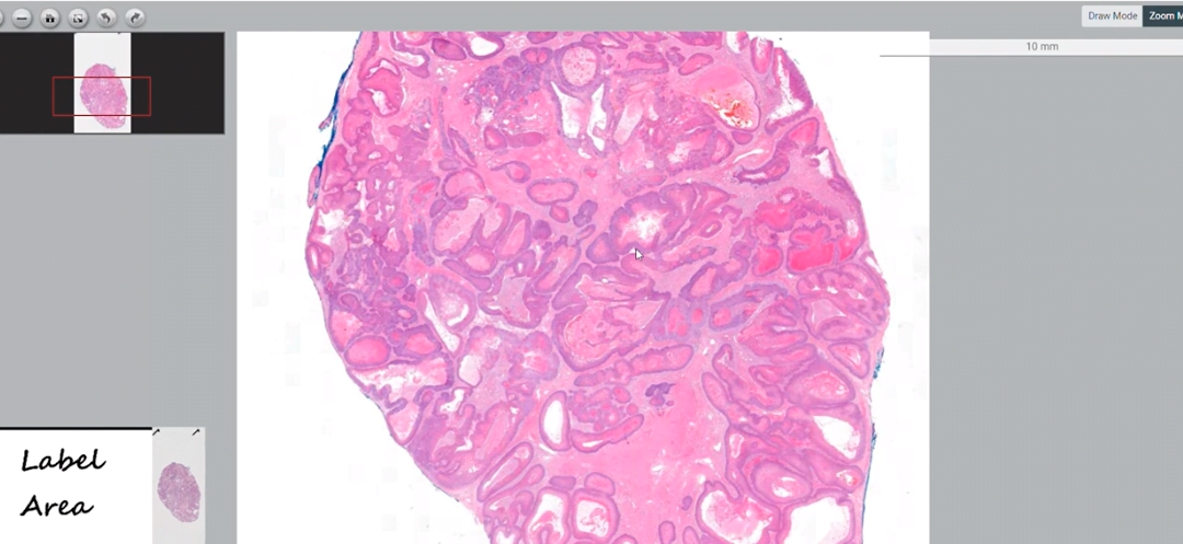
In this episode of Digital Dermatopathology Digest, Rajni Mandal, MD, dermatopathologist at PathologyWatch, explains the characteristics of proliferating pilar cysts.
“Proliferating pilar cysts are most commonly seen in adult women in the scalp,” Mandal says. “Most cases arise from a pre-existing pilar cyst, due to unknown triggers.”
In the video, Mandal goes on to identify the defining features of two categories of proliferating pilar cysts:
Benign Proliferating Pilar Cyst
Cyst epithelium proliferates within the center of the cyst, giving it a “rolls and scrolls” appearance. The proliferating cells are trichilemmal, with small nuclei having smooth nuclear contours and a uniform chromatin pattern. The cyst lining has no granular layer, with abrupt dense, compact, pink keratin formation.
Malignant Proliferating Pilar Cyst
Also known as trichilemmal carcinoma, malignant proliferating pilar cysts differ from the benign version in key ways. In addition to the above characteristics, malignant cysts show cell crowding and cellular atypia (i.e., nuclei of varying size and shape). These cysts display mitotic activity, infiltrative growth, and cytologic atypia.
Whether you’re in residency, studying for board exams, or a practicing dermatologist looking to stay sharp, the Digital Dermatopathology Digest video series is your informational and convenient source for dermatopathology review. Find the full series here.

